BH Breast Health Studio Research
Models Frozen
⚠️ Research / Educational Prototype · Not for clinical diagnosis

Advancing Breast Health Intelligence Through Transparent AI Research.

An open academic platform unifying classical fine needle aspirate morphometry, deep learning mammography analysis, empirical calibration, and transparent explainability for oncology research and public health education.

FROZEN RESEARCH RUNTIME

Validated against retrospective cohorts (WDBC & CBIS-DDSM). Model parameters and decision boundaries cryptographically verified.

CBIS-DDSM TEST CASE #1042 EFFICIENTNET-B0 INFERENCE
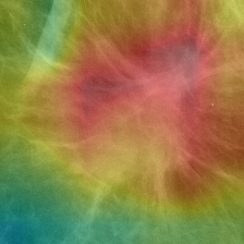
CBIS-DDSM mammography full processed image with Grad-CAM coarse attention overlay
Grad-CAM Coarse Attention Conv5 Feature Saliency (Not Tumor Boundary)
Study A · WDBC (FNA) Logistic Regression
ROC-AUC 99.54%
Raw Cutoff ≥ 0.360
Study B · CBIS-DDSM EfficientNet-B0
ROC-AUC 72.29%
Raw Cutoff ≥ 0.515
Academic Mission

Bridging rigorous machine learning, reproducible medicine, and public breast health literacy.

Contemporary healthcare AI often operates as an opaque black box, obscuring the critical distinction between statistical probability and clinical diagnosis. Our mission is to dismantle that opacity through transparent model architectures, honest calibration reporting, and traceable, peer-reviewed educational literature.

By decoupling distinct biological modalities—cellular morphometry from digital radiography—we respect the distinct clinical questions each modality addresses while illustrating where machine learning can support, rather than replace, human expertise.

Scientific Evidence & Methodological Transparency

The Research Story: Two Separate Studies, One Commitment to Rigor

Understand the project in 60 seconds: Study A and Study B investigate fundamentally different clinical diagnostic domains using completely separate modalities and patient cohorts. Candidates were evaluated strictly on development/validation evidence — never final test sets.

⚠️ Critical Scientific Guardrail: WDBC evaluates high-resolution cytological aspirates from palpable lesions; CBIS-DDSM evaluates screening mammograms with variable tissue densities. No cross-study ranking or combined leaderboards exist. Evaluations are strictly within-study.
Step A · What Did We Study?

Two Distinct Biological Modalities

Study A: 569 FNA biopsy cases with 30 nuclear morphology features (WDBC).
Study B: 1,696 digital mammography scans (CBIS-DDSM full processed images).

Step B · What Did We Test?

Competitive Model Candidates

Study A: Logistic Regression (selected), Random Forest, XGBoost.
Study B: EfficientNet-B0 (selected), ResNet50, Custom CNN.

Step C · How Did We Select?

Validation-First Selection

Candidates were selected strictly on development/validation splits. Held-out test sets remained locked until post-selection confirmation.

Study A · Classical ML 569 Cases · 30 FNA Features

WDBC Cytology Selection

Candidate screening protocol: 5-Fold Out-Of-Fold (OOF) Cross-Validation on development data.

Development Evidence (5-Fold OOF CV)
Candidate ROC-AUC PR-AUC Sens. Spec. Bal. Acc Brier Status
Logistic Regression 0.9950 0.9941 97.65% 96.84% 97.24% 0.0200 Selected
Random Forest 0.9876 0.9859 95.88% 96.49% 96.19% 0.0305 Evaluated
XGBoost 0.9939 0.9924 95.29% 98.95% 97.12% 0.0225 Evaluated
↓ Development Selection: Logistic Regression

Why Logistic Regression?

Logistic Regression demonstrated the strongest, most stable diagnostic balance across all five development folds, providing top-tier discriminatory power while remaining fully interpretable with direct coefficient weights.

  • Development-only selection: Selected strictly on 5-fold cross-validation evidence prior to evaluating test data.
  • Highest ROC-AUC (0.9950) & PR-AUC (0.9941): Outperformed ensemble tree alternatives in discriminatory accuracy.
  • Superior probability calibration: Lowest Brier score (0.0200) and highest balanced accuracy (97.24%).
  • Convex & robust: L2 regularization prevents overfitting on small clinical cohorts (569 cases) compared to non-linear splits.
↓ Locked Test Report (Post-Selection Verification)
Final Held-Out Report WDBC Test Split (114 Cases)
Frozen decision rule: raw malignant probability ≥ 0.360
Accuracy 97.37%
ROC-AUC 0.9954
PR-AUC 0.9932
Sensitivity 95.24%
Specificity 98.61%
Balanced Acc. 96.92%
TN: 71 (Benign) FP: 1 FN: 2 TP: 40 (Malignant)
Post-selection verification report. Held-out test data did NOT drive candidate selection.
Inspect Study A Evidence Figures (ROC Curves & SHAP)
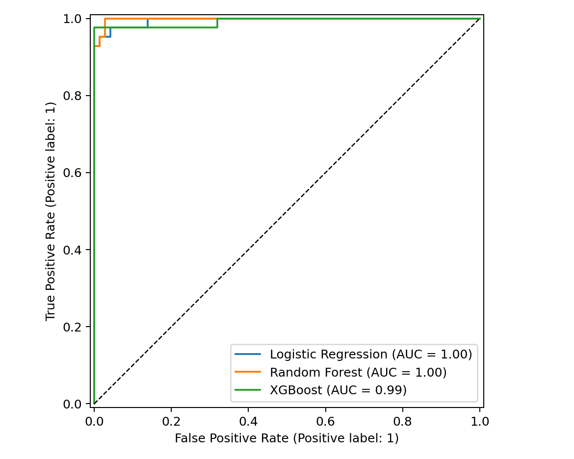
Study A ROC curves comparison for Logistic Regression, Random Forest, and XGBoost
ROC curves comparing cross-validation discriminatory trajectories across classical ML candidates.
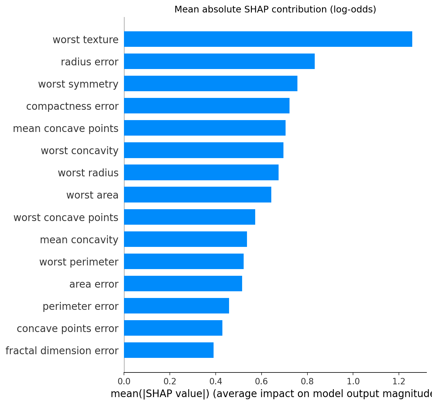
SHAP global feature importance bar plot for WDBC Logistic Regression
SHAP global feature attribution validating primary nuclear contour dependencies.
Study B · Deep Learning Full Processed Mammography

CBIS-DDSM Vision Selection

Candidate screening protocol: Validation-First Evidence on frozen validation split.

Validation-First Evidence (Validation Split)
Candidate Cutoff ROC-AUC PR-AUC Sens. Spec. Bal. Acc Brier Status
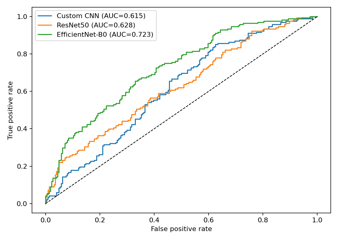
EfficientNet-B0 0.515 0.7044 0.6152 68.13% 63.48% 65.80% 0.2327 Selected
ResNet50 0.470 0.6690 0.5733 80.00% 47.83% 63.91% 0.2349 Evaluated
Custom CNN 0.500 0.6373 N/A 51.88% 70.43% 61.15% 0.2468 Evaluated
↓ Validation-First Selection: EfficientNet-B0

Why EfficientNet-B0?

EfficientNet-B0 achieved the highest validation discrimination while maintaining a balanced trade-off between sensitivity and specificity, avoiding the extreme specificity collapse observed in heavier architectures.

  • Validation-first selection: Candidate selection occurred strictly on the validation split before evaluating held-out test data.
  • Highest validation ROC-AUC (0.7044) & PR-AUC (0.6152): Outperformed ResNet50 and Custom CNN on unseen scans.
  • Lowest validation calibration error: Achieved lowest Brier score (0.2327) and highest balanced accuracy (65.80%).
  • Avoided false-positive surge: ResNet50 specificity collapsed to 47.83%, whereas EfficientNet-B0 maintained 63.48% specificity.
↓ Locked Test Report (Post-Selection Verification)
Final Held-Out Report Full Processed Test Split (392 Scans)
Frozen decision rule: raw malignant probability ≥ 0.515
Accuracy 64.80%
ROC-AUC 0.7229
PR-AUC 0.6564
Sensitivity 67.86%
Specificity 62.50%
Brier Score 0.2297
TN: 140 (Benign) FP: 84 FN: 54 TP: 114 (Malignant)
Post-selection verification report. Held-out test data did NOT drive candidate selection.
Inspect Study B Evidence Figures (ROC Curves & Grad-CAM)
Study B ROC comparison for transfer learning mammography models
ROC curves comparing validation and test trajectories for mammography deep learning backbones.
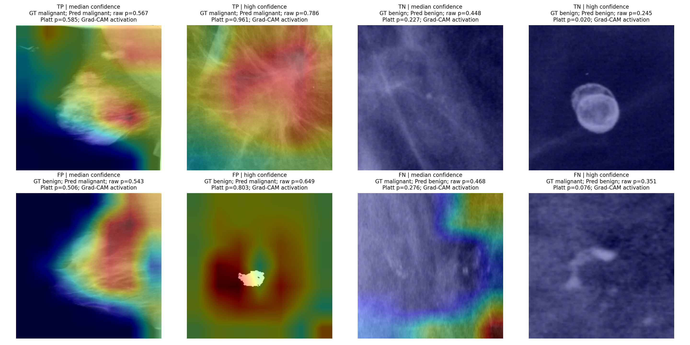
Grad-CAM heatmaps for EfficientNet-B0 mammography classification
Grad-CAM visual attribution highlighting focal radiologic density regions.
Academic Research Dossiers

Explore Technical Research Pages

For researchers and clinical investigators seeking comprehensive experimental protocols, distribution splits, bootstrap confidence intervals, and explainability maps.

Decision Rule & Telemetry Semantics

From Probability to Prediction

Explore how raw model probabilities are compared with frozen research decision thresholds to produce a research prediction. For the mammography model, a separate Platt calibrated probability is shown for reliability interpretation only.

🔬 Research Explainer: How does the frozen model turn a continuous probability into a categorical research prediction?
SELECT DEMONSTRATION SAMPLE:
Interactive demonstration sample (WDBC Cytology)
Fine Needle Aspirate (FNA) 30 Nuclear Morphometry Features
Raw Malignant Probability Model Output · Classification Basis
88.4%
0.0 (Benign) Frozen research decision threshold: ≥ 0.360 1.0 (Malignant)
Margin from threshold: +52.4% above cutoff

This raw probability is the direct mathematical output evaluated against the frozen research decision threshold to produce the categorical prediction.

Decision Rule Architecture Study A Runtime Contract
Raw Retained
if raw malignant probability ≥ 0.360:
  → Malignant Prediction
else:
  → Benign Prediction
Frozen Decision Boundary: 0.360

No post-hoc Platt scaling is applied to Study A. The frozen Logistic Regression runtime retains its raw sigmoid probability directly for decision assignment and research evaluation.

Evaluated Model Candidate: Logistic Regression (30 FNA Features)
Malignant Prediction (Raw probability 0.884 ≥ 0.360 threshold)
Evidence-Based Public Health Literacy

Breast Health Education & Clinical Guidance Hub

An accessible, clinically sourced knowledge resource for patients, families, and researchers. Explore breast biology, evidence-based screening schedules, early warning signs, nutrition, survivorship care, and curated clinical video lectures.

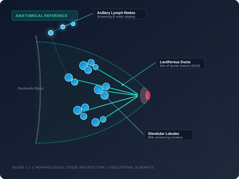
Anatomical illustration of female breast tissue showing milk ducts, lobules, and adipose tissue
Medical Foundations

Understanding Breast Tissue & Cellular Architecture

The adult female breast consists of glandular tissue (lobules and milk ducts), fibrous connective stroma, and protective adipose (fatty) tissue. Most breast malignancies originate within the epithelial lining of the terminal ductal lobular units (ductal carcinoma) or the milk-producing lobules (lobular carcinoma).

Recognizing subtle structural changes—such as microcalcifications, asymmetrical densities, or cellular pleomorphism—is the core objective of radiological screening and cytological biopsy analysis.

Ductal vs Lobular Stroma & Adipose Terminal Duct Lobular Units (TDLU)
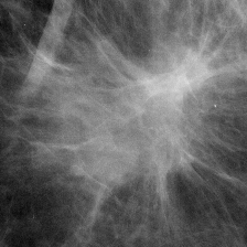
Clinical high-resolution mammogram demonstrating focal soft-tissue asymmetry
Diagnostic Imaging

Screening Guidelines & Mammography Modalities

Screening mammography remains the primary evidence-based modality proven to reduce breast cancer mortality through early detection before palpable symptoms appear.

Breast tissue density (categorized under BI-RADS A through D) significantly influences radiographic sensitivity. High tissue density can obscure lesions, leading clinicians to recommend supplementary automated breast ultrasound (ABUS) or breast MRI.

BI-RADS Staging Dense Tissue Sensitivity Digital Tomosynthesis

Evidence-Based Screening Schedules by Organization

Separated Evidence Sources

The American Cancer Society (ACS) and U.S. Preventive Services Task Force (USPSTF) publish distinct, evidence-based recommendations. They are presented separately below to maintain methodological rigor without conflating recommendations.

ACS Guidance American Cancer Society Recommendations (Average Risk)
Source: Oeffinger et al., JAMA 2015;314(15):1599-1614 (ACS Guidelines)
Age Group ACS Recommendation Frequency Clinical Shared Decision
Ages 40 – 44 Women have the choice to begin annual screening mammography based on individual preference and discussion with their clinician. Annual (Elective) Optional early initiation
Ages 45 – 54 Women should undergo regular screening mammography. Peak evidence-backed benefit for annual detection. Every Year Recommended routine screening
Ages 55 and Older Women can transition to biennial (every 2 years) screening or continue annual screening, continuing as long as overall health is good and life expectancy is 10+ years. Every 1 to 2 Years Continue while in good health
USPSTF Guidance U.S. Preventive Services Task Force (2024 Final Recommendation)
Source: USPSTF Recommendation Statement (JAMA 2024)
Population USPSTF Recommendation Interval Evidence Grade
Women Aged 40 – 74
Average Risk
Biennial screening mammography is recommended for all average-risk women starting at age 40 to balance early mortality reduction against overdiagnosis. Every 2 Years (Biennial) Grade B (Moderate Net Benefit)
Women Aged 75 and Older Current published evidence is insufficient to assess the balance of benefits and harms of screening mammography. Individualized Grade I (Insufficient Evidence)
Dense Breasts (BI-RADS C/D) Current evidence is insufficient to assess the balance of benefits and harms of supplemental screening (ultrasound or MRI) beyond standard mammography. Per Physician Assessment Grade I (Insufficient Evidence)
High-Risk Protocol Consideration (ACR / ACS / NCCN) Genetics & Radiation History

Women with a known BRCA1/BRCA2 mutation, a first-degree relative with a mutation, a calculated lifetime breast cancer risk ≥ 20%, or prior mantle chest radiation therapy between ages 10 and 30 are recommended to begin annual contrast-enhanced breast MRI and screening mammography starting at age 30 under specialist clinical oversight.

Source Registry Citations: American Cancer Society (Oeffinger et al., JAMA 2015;314(15):1599-1614); U.S. Preventive Services Task Force (JAMA 2024;331(22):1918-1930); American College of Radiology Appropriateness Criteria (2023).
Early Recognition

Recognizing Warning Signs: When to Seek Medical Evaluation

Familiarity with your baseline breast anatomy is vital for noticing changes early. The following visual guide outlines common changes that warrant a timely professional clinical evaluation.

ℹ️
Important Educational Framing: These changes can have many non-malignant causes (such as benign cysts, fibroadenomas, hormonal fluctuations, or benign infections). Experiencing one of these signs does not mean you have breast cancer, but persistent or concerning changes should always be evaluated promptly by a qualified healthcare professional.
🔍

New Lump or Mass

A distinct lump in the breast or armpit. Malignant lumps are frequently painless and hard with irregular borders, but tender, soft, or rounded lumps can also occur.

🩺

Swelling or Thickening

Swelling of all or part of the breast, or an area of thickened tissue that feels noticeably different from surrounding areas, even without a distinct lump.

🪞

Skin Dimpling or Puckering

Surface indentations, puckering, or texture changes resembling an orange peel (peau d'orange), often caused by subtle tethering of subcutaneous ligaments.

🔄

Nipple Retraction or Inversion

A nipple that newly turns inward, inverts, or shifts orientation. Pre-existing lifelong inverted nipples are generally benign, but recent changes require review.

🔴

Redness, Flaking, or Scaliness

Persistent redness, flaking, scabbing, or thickening of the nipple or areolar skin that does not resolve with standard topical moisturizers.

💧

Unusual Nipple Discharge

Discharge occurring spontaneously without squeezing, particularly if clear, watery, or blood-tinged, and localized to one breast.

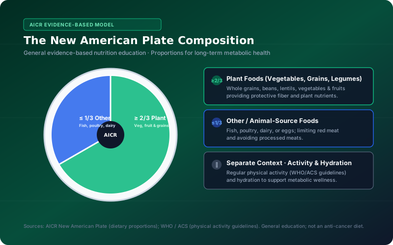
AICR New American Plate proportion model showing plant-forward dietary pattern and hydration
Public Health & Prevention

Evidence-Based Nutrition & Lifestyle Modalities

Epidemiological evidence compiled by the American Institute for Cancer Research (AICR), World Cancer Research Fund (WCRF), and Memorial Sloan Kettering (MSK) establishes that balanced dietary patterns support long-term wellness and survivorship.

The AICR New American Plate model recommends covering at least 2/3 of your plate with plant foods (vegetables, fruits, whole grains, and legumes/beans), with 1/3 or less dedicated to other or animal-source foods (such as fish, poultry, or dairy).

As a distinct lifestyle recommendation, the World Health Organization (WHO) and American Cancer Society advise regular physical activity (150–300 minutes weekly) to support metabolic health.

AICR New American Plate (≥ 2/3 Plant) MSK Survivorship Education Active Treatment vs Prevention
ℹ️
Evidence-Based Sourcing Notice: Nutritional guidance supports overall health but does not cure cancer. No specific food cures cancer or lowers mathematical AI risk scores. Nutritional needs during active chemotherapy/radiation differ substantially from general prevention and must be managed directly by clinical oncology dietitians (e.g., MSK Evelyn H. Lauder Breast Center protocols).
Survivorship & Self-Care

Supportive Care, Recovery & Survivorship Pathways

Navigating clinical investigations or recovering after treatment involves emotional resilience, physical pacing, and coordinated family support.

🌙

Rest & Energy Pacing

Acknowledge fatigue as a physiological signal. Prioritize sleep, take short restorative breaks, and delegate non-essential daily tasks during treatment.

🚶‍♀️

Gentle Movement

Doctor-approved walking or range-of-motion arm exercises help sustain stamina, manage lymphedema risk, and boost mood during recovery.

📝

Symptom & Health Diary

Keeping a brief log of symptoms, medication schedules, and questions empowers productive, focused dialogues during subsequent clinic visits.

🤝

Emotional & Family Support

Engage oncology social workers, peer support groups, and family caregivers. Open communication relieves anxiety and reduces isolation.

Clarity & Evidence

Separating Common Myths from Medical Facts

Misinformation can produce unwarranted panic or delay essential medical care. Here is the verified clinical evidence behind common misconceptions.

❌ Common Myth

"Finding a breast lump always means you have cancer."

✅ Medical Fact

Approximately 80% of breast lumps evaluated in clinical practice turn out to be benign conditions, such as fluid-filled cysts or benign fibroadenomas. However, any new lump must be professionally examined to establish an accurate diagnosis.

❌ Common Myth

"If breast cancer does not run in my family, I am not at risk."

✅ Medical Fact

Roughly 85% of breast cancers occur in individuals with no prior family history. They arise primarily from non-inherited factors, aging, hormonal exposure, and somatic genetic mutations occurring over a lifetime.

❌ Common Myth

"An AI algorithm or online tool can diagnose breast cancer."

✅ Medical Fact

AI software calculates statistical probability estimates based on retrospective research data. Only a licensed physician using physical exams, diagnostic mammography/ultrasound, and pathology biopsy can provide a definitive medical diagnosis.

❌ Common Myth

"Screening mammograms expose patients to dangerous radiation levels."

✅ Medical Fact

Screening mammograms utilize very low, tightly regulated radiation doses—comparable to roughly 7 weeks of natural background radiation. The proven mortality reduction benefit of early detection far outweighs the minimal radiation exposure.

Verified Video Education

Curated Clinical Video Library

Educational video lectures and clinical explainers curated from verified oncology institutions. Videos use lazy loading to preserve page performance until activated.

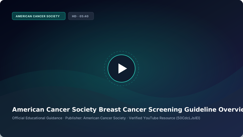
ACS Breast Cancer Screening Guideline Overview Video Poster
05:40 min
Screening Protocols

American Cancer Society Screening Guideline Overview

An authoritative walkthrough outlining evidence-based screening recommendations, age milestones, annual mammography schedules, and dense breast tissue clinical context.

Publisher: American Cancer Society (ACS)
Duration: 05:40 min · Verified Official YouTube (50CdcLJsIEI)
Topic: Screening Guidelines & Clinical Mammography
Radiographic scan illustrating screening early detection
02:21 min
Clinical Evidence

The Science Behind Breast Cancer Screening Guidelines

Discussion of clinical evidence, mortality reduction modeling, and risk-benefit analyses guiding regular screening intervals and clinical practice.

Publisher: American Cancer Society (ACS)
Duration: 02:21 min · Verified Official YouTube (oZYRmApgoUI)
Topic: Interval Evidence & Mortality Reduction
Practical Q&A

Frequently Asked Questions

Clear answers to common questions regarding AI probability scores, screening modalities, cytology features, and analysis privacy.

What does an AI probability score mean?
An AI probability score represents a mathematical estimate generated by comparing your input features against retrospective clinical study cohorts. For example, Study A evaluates 30 cytological features against the frozen 0.360 threshold. The score indicates statistical pattern alignment, not a clinical diagnosis or medical certainty.
Does this platform diagnose breast cancer?
No. This software is developed strictly as an academic research and educational prototype (clinical_use = false). It cannot provide a definitive diagnosis. Definite medical diagnosis requires physical examination, diagnostic imaging, and histological biopsy interpreted by a licensed pathologist and oncologist.
What is screening mammography and why does tissue density matter?
Screening mammography uses low-dose X-rays to image the internal architecture of breast tissue. When tissue is categorized as dense (BI-RADS C or D), both fibroglandular tissue and tumors appear white on radiography, potentially masking small lesions. In such cases, supplemental ultrasound or MRI may be recommended by your clinician.
What are WDBC fine needle aspirate (FNA) nuclear features?
The Wisconsin Diagnostic Breast Center (WDBC) dataset contains 30 computerized morphometry measurements derived from digitized images of fine needle aspirates. These capture nuclear characteristics such as mean radius, texture, perimeter, area, smoothness, compactness, concavity, concave points, symmetry, and fractal dimension.
Why might the cytology (ML) and mammography (DL) models produce different results?
Study A (WDBC) and Study B (CBIS-DDSM) evaluate completely different biological modalities from separate patient cohorts. Cytology evaluates microscopic nuclear morphology of aspirated cells from a focal biopsy, whereas mammography evaluates gross radiological tissue patterns across an entire breast image. Disagreements reflect differing diagnostic domains, not algorithmic conflict.
What should I do if I notice a concerning change or receive an elevated AI result?
Do not panic. Schedule a clinical consultation with your primary care physician or a specialized breast health specialist. Bring previous mammography films or medical records if available. Most breast symptoms have benign causes, but prompt clinical evaluation ensures accurate clarity and peace of mind.
Where is my analysis history saved and how is my data protected?
When signed in, your analysis history is stored securely in the system database linked to your user account. Guest explorations do not store personal records. For healthcare providers, the Doctor Workspace isolates records under designated patient IDs. Data is never shared with third parties or used for external advertising.
Does this educational content replace a doctor's consultation?
Absolutely not. All educational articles, screening tables, and symptom guides are provided solely for public health literacy and to help patients prepare for productive conversations with their healthcare team. Never delay or modify medical treatment based on online educational content.
Safety & Regulatory Framework

Critical Research Limitations & Ethical Guardrails

This software is developed strictly as an academic research and educational prototype. It does not constitute a certified medical device and must never be used as a substitute for professional medical advice, diagnosis, or clinical management.

  • No Clinical Clearance: This platform is not cleared by the FDA, CE, or regional health authorities for diagnostic usage (clinical_use = false).
  • Pathology Ground Truth Required: Computer vision and machine learning inferences do not replace microscopic histopathological examination or clinical palpation.
  • Retrospective Training Cohorts: Models were trained on retrospective public datasets (WDBC & CBIS-DDSM) with demographic limitations and without prospective multi-site clinical trials.
  • Unpaired Multimodal Heuristic: The multimodal demo employs an unvalidated 40/60 weighted software heuristic; no clinically matched patient cohorts were evaluated simultaneously.
  • Demo Mammogram Provenance & Attribution: Demo mammograms are derived from the Curated Breast Imaging Subset of DDSM (CBIS-DDSM) hosted on The Cancer Imaging Archive (TCIA). Production distribution licensing remains a documented PRE-DEPLOY BLOCKER.
Academic Authorship

Academic Research Team

Conducted within the School of Information and Communication Technology under faculty academic supervision.

ND
Nguyễn Bá Duy Machine Learning & WDBC Study
TA
Trần Mỹ Anh Deep Learning & Computer Vision
HA
Hoàng Nhật Anh Model Calibration & Metrics
GH
Nguyễn Huy Giang Systems & Platform Architecture
TD
Ngô Tiến Đạt Explainability & Evaluation
Project Supervisor: GV. Đoàn Thị Thanh Hằng
Get Started

Explore the Breast Health Intelligence Studio

Test model predictions, examine SHAP and Grad-CAM interpretability visualizations, and review frozen research checkpoints.

Start AI Analysis Review Research Story